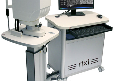
Funduskamera z optyką adaptywną RTX-1
Ultranowoczesna funduskamera z zastosowaniem optyki adaptywnej pozwala na uzyskanie rozdzielczości obrazu na poziomie 3-4 µm, co umożliwa obserwcację takich struktur jak:
- czopki,
- lamina cribrosa,
- mikronaczynia siatkówki włącznie z pomiarem grubości ich ścian a także analizą WLR (wall to lumen ratio).
Więcej informacji na stronie producenta: http://www.imagine-eyes.com
Zdjęcia produktu
Materiały informacyjne
Artykuły
- M RCC 003 a4.3 – DR – XV-XX
- M RCC 005 a4.3 – Macular hole – Besancon
- M RCC 007 a4.3 – Macular hole – VRMNY Zespół pomarszczenia plamki
- M RCC 010 a4.3 – GA foveal sparing- XV-XX
- M RCC 011 a4.3 – GA- XV-XX
Literatura
- 2010.11 Veterinary Ophthal – Rosolen – AO imaging in feline retina
- 2011.02 Archiv Ophth. – Audo Nakashima Paques – Poppers toxicity with AO images
- 2013.01 Clin Ophth – Hayashi – AO fundus Images of cones in RP
- 2013.01 Sensors – Lombardo – AO for retinal imaging
- 2013.03 OPO – Lombardo – Eccentricity dependent changes of density, spacing and packing arrangement of parafoveal cones
- 2013.03 Retina – Lombardo – Analysis of Retinal Capillaries in Type 1 Diabetes and DR using AO
- 2013.03 Retina – Lombardo – Interocular Symmetry of Parafoveal Cone Density Distribution
- 2013.04 IOVS – Gocho Paques – AO imaging of GA
- 2013.05 Clin Ophth – Hayashi – Analysis of macular cones in OMD
- 2013.07 BioMed OE – Lombardo – Sample window size and orientation influence on cone density
- 2013.08 Jama – K Bailey Freund – Paracentral Acute Middle Maculopathy
- 2013.08 Jama – Mrejen – APMPPE as Choroidopathy and AO
- 2013.10 Ophthalmology – Mrejen, Spaide – PR mosaic in pseudo and soft drusen with AO
- 2013.11 BioMed Res Int – Gocho – Mycrosystic edema rtx1
- 2013.12 Doc Ophthalmol – Faure Gocho Audo – AO imaging in ocular siderosis
- 2014.01 Indian J Ophthal – Battu Dabir – AO retinal imaging
- 2014.01 Ophthalmology – Da Cruz – AO Img shows Rescue of Macula PR’s
- 2014.02 Ophthalmology – Errera Paques – AO imaging in vasculitis – in Press
- 2014.02 Saudi Journal of Opht – Kozak – Imaging retina with AO
- 2014.03 BioMed OE – Ramaswamy Lombardo Devaney – Registration AO RNFL images
- 2014.03 PlosOne – Obata Yanagi – AO imaging in early AMD
- 2014.03 Retina – Lombardo – AO imaging cones type 1 diabetes
- 2014.04 Br J Ophthalmol – Muthiah – cone analysis rtx1
- 2014.04 CEO – Saleh – Reliability cone counting with rtx1
- 2014.04 J Hypertens – Koch et al – AO imaging in HT 2014.06 Br J Ophthal-Saleh-Cone loss after retinal detachment
- 2014.07 Acta Ophth – Bek – AO in diabetic retinopathy
- 2014.07 Retina – Paques et al – AO imaging Gunn dots – v_proof
- 2014.09 J Ophth – Gocho Kameya – AO in Bietti disease
- 2014.09 Plos One – Lombardo – AO technical factors
- 2014-10 Eye – Dabir et al – Cone density vs eccentricity
- 2015.01 Am J Opht – Jacob et al – Meaning of visualizing cones with AO
- rtx1 – list of publications

